Figure 1.
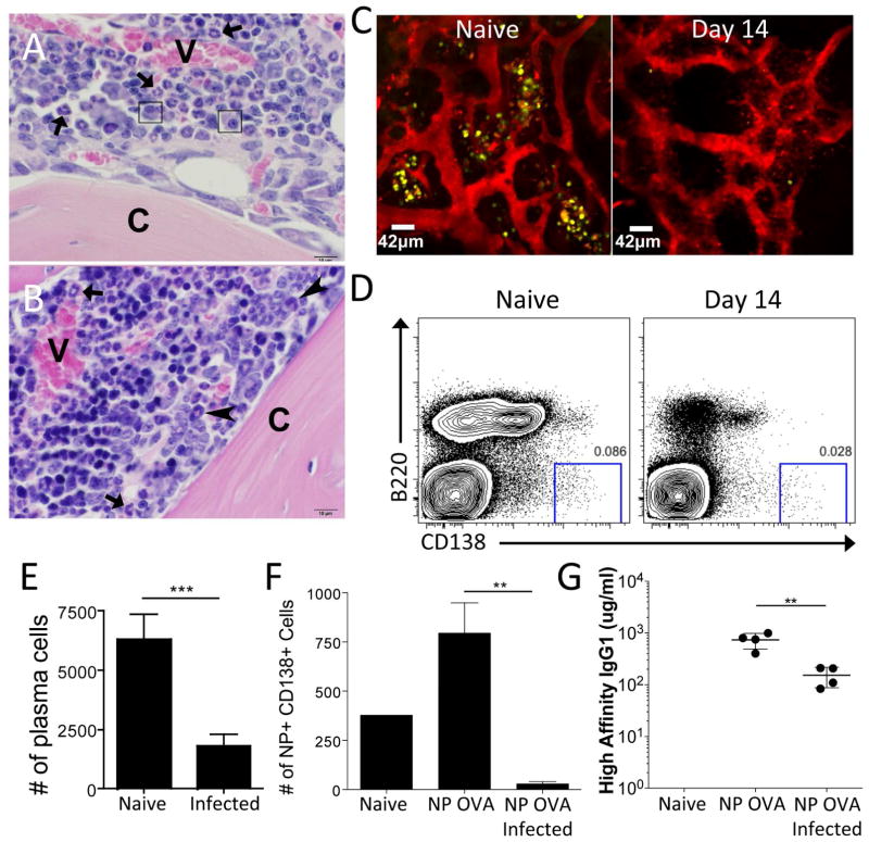
Acute Toxoplasma gondii infection results in a loss of PC in the BM. (A) Naïve mouse. The marrow cavity contains vascular sinuses (V) surrounded by mature neutrophils (arrowheads) admixed with predominantly myelopoietic precursors and few mature PC (arrows). Bone cortex (C). (B) Day 14 T. gondii infected mouse. Medullary vascular sinuses (V) are surrounded by increased numbers of hematopoietic progenitors characterized by hyperchromatic nuclei. Few mature neutrophils (arrowheads) and immature band neutrophils (arrows) are observed. Mature PC are not identified. Bone cortex (C). (C) Naïve or infected BLIMP1-YFP reporter mice were imaged using intravital 2-photon microscopy of the skull BM. The BLIMP1-YFP-expressing cells are yellow and quantum dots were injected intravenously to label the vasculature red. At least 3 mice were imaged for each time point. (D–E) BM from naïve or infected WT mice was evaluated by flow cytometry (using a “dump” gate to eliminate CD3+, F4/80+, and/or Gr1+ potential contaminating cells) for the presence of PC. (F–G) WT mice were immunized with NP-OVA. 6 weeks later, these mice were challenged with T. gondii and evaluated for NP-specific PC (F) and NP-specific antibody titers (G).

