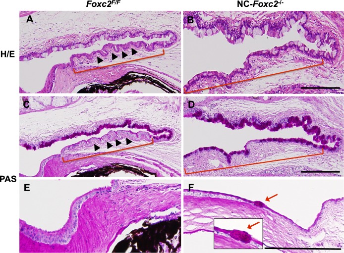
Figure 5.
Ectopic goblet cell formation in the corneal epithelium of adult NC-Foxc2−/− mice. (A–F) H/E (A, B) and PAS (C–F) stain of eyes in control (Foxc2F/F) and NC-Foxc2−/− mice at 8 weeks. Periodic acid Schiff stain was performed to detect mucin-secreting goblet cells. Neural crest-Foxc2−/− mice had defective stratification of columnar epithelium (B, D, brackets) in conjunctiva, compared with normal stratified columnar shape without goblet cells (A, C, brackets, arrowheads) in the conjunctival epithelium of control mice. Neural crest-Foxc2−/− mice displayed conjunctival goblet cell expansion (B, D, brackets) and an ectopic goblet cell (F, red arrows, inset) in the peripheral corneal epithelium. Scale bars: 100 μm.

