
Figure 3.
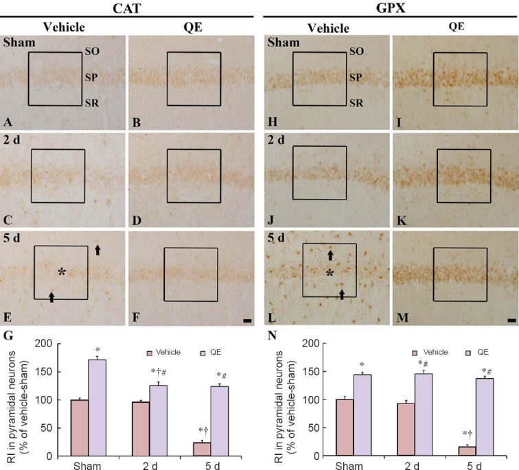
CAT and GPX immunoreactivities in the hippocampal CA1 region of the vehicle-sham (A, H), vehicle-ischemia (C, E, J, L), QE-sham (B, I) and QE-ischemia (D, F, K, M) groups.
At 2 days post-ischemia, CAT and GPX immunoreactivities were gradually decreased in the hippocampal CA1 pyramidal neurons and hardly detected 5 days post-ischemia (asterisks); however, non-pyramidal cells (arrows) show CAT and GPX immunoreactivities. In the QE-sham group, CAT and GPX immunoreactivities were significantly increased in the hippocampal CA1 pyramidal neurons, and in the QE-ischemia group, CAT and GPX immunoreactivities were also strong at 2 and 5 days post-ischemia. Scale bars are 20 μm in F and M, valid for A–M. (G, N) Relative immunoreactivity (RI) as percent of CAT and GPX in hippocampal CA1 pyramidal neurons in a 140 × 140 μm2 (boxes). n = 7 at each time point in each group.*P < 0.05, vs. vehicle-sham group; †P < 0.05, vs. respective pre-time point group; #P < 0.05, vs. corresponding vehicle-ischemia group. Data in F and M are expressed as the mean ± SEM. CAT: Catalase: GPX: glutathione peroxidase; QE: quercetin; SO: stratum oriens; SP: stratum pyramidale; SR: stratum radiatum.