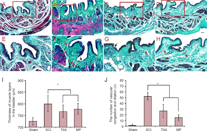
Figure 4.
Effect of TIIA on histology of bladder tissue at 4 weeks after SCI (Masson's trichrome staining).
(A, E) Normal bladder morphology in sham group: The red color indicates muscle fibers, and the green color indicates collagen. (B, F) Bladders from SCI group reveal the presence of edema (pink) and proliferation of urothelial layers. The continuity of the umbrella cell layer of the urothelium is disrupted. A marked neutrophil infiltration to the suburothelial tissue as well as blood vessel congestion and dilation are observed in the TIIA (C, G) and MP (D, H) groups. The urothelium is disrupted partially and some blood vessel congestion and dilation can be seen. Scale bars: 100 μm for A–D; 50 μm for E–H. The disrupted urothelium is shown by black arrows. Vascular congestion and dilation are shown by red arrows. (I) The thickness of muscle layers. (J) The number of vascular congestion and dilation (n = 8 in each group; *P < 0.05). SCI: Spinal cord injury; TIIA: tanshinone IIA; MP: methylprednisolone.

