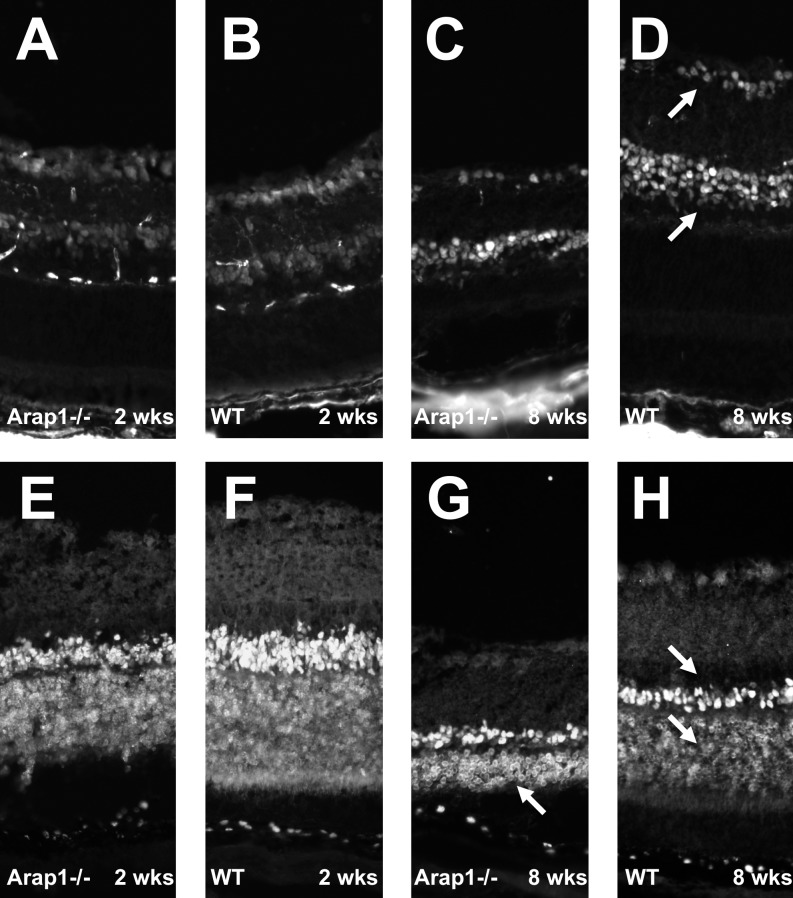
Figure 4.
Arap1−/− mice form appropriate numbers and types of laminated retinal neurons. Immunohistochemistry using anti-Pax6 was performed to mark retinal ganglion cells and amacrine cells (arrows in [D]). Arap1−/− retina from 2 weeks postnatal mice (A) showed a similar staining pattern as wild-type (B) animals at this age. Anti-Otx2 labeling was used to mark bipolar and photoreceptor cells (arrows in [H]). A similar pattern of Otx2 staining was seen between Arap1−/− (E) and wild-type (F) controls at 2 weeks postnatal age. By 8 weeks of age, the Pax6 labeling in the degenerating Arap1−/− retina (C) remained similar to controls (D); however, the Otx2-labeled photoreceptor layer was markedly thin by 8 weeks in Arap1−/− mice (arrow, G) when compared with wild-type controls (lower arrow, H).

