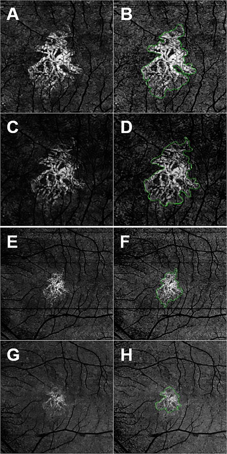
Figure 1.
En face SS- and SD-OCTA images from the left eye of a 70-year-old woman with CNV secondary to AMD. All images were processed from the corresponding volumetric datasets using the same algorithms that were applied to a slab that extended from the outer retina to the choriocapillaris and included the removal of the projection artifacts from the retinal vasculature. (A) Spectral-domain OCTA 3 × 3-mm2 scan. (B) Spectral-domain OCTA 3 × 3-mm2 scan with an outline of the CNV and an area of 1.51 mm2. (C) Spectral-domain OCTA 3 × 3-mm2 scan. (D) Spectral-domain OCTA 3 × 3-mm2 scan with an outline of the CNV and an area of 1.51 mm2. (E) Spectral-domain OCTA 6 × 6-mm2 scan. (F) Spectral-domain OCTA 6 × 6-mm2 scan with an outline of the CNV and an area of 1.52 mm2. (G) Spectral-domain OCTA 6 × 6-mm2 scan. (H) Spectral-domain OCTA 6 × 6-mm2 scan with an outline of the CNV and an area of 1.13 mm2. In this example, the area measurements were similar for the 3 × 3-mm2 scans, but SS-OCTA imaging showed a larger lesion for the 6 × 6-mm2 scans.

