
Figure 1.
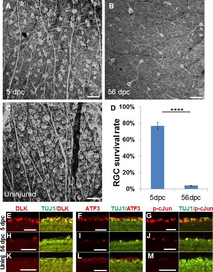
Axotomy-induced cell death and injury-signal proteins at acute and chronic time points. (A–B) β III tubulin immunoreactive (i.e., TUJ1 antibody immunostained) whole-mount retinae at 5 days (A) and 56 days (B) after optic nerve crush, and uninjured control retina (C). (D) Quantification of RGC survival, normalized to uninjured contralateral retina (n = 5/group, ****P = 0.0000026, Student's unpaired t-test). (E–M) Retinal section images of immunohistochemical analysis for DLK, ATF3, p-c-Jun signals in the ganglion cell layer of mice 5 days (E–G) and 56 days (H–I) after injury and without injury (K–M) (n = 5/group). Error bars: SEM. Scale bars: 50 μm.