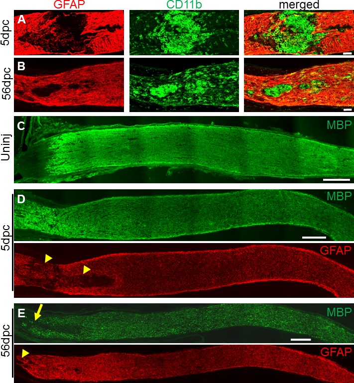
Figure 2.

Changes in lesion and its penumbra within acutely and chronically injured optic nerves. (A–B) Glial fibrillary acidic protein and CD11b immunostained lesion sites at 5 days (A) and 56 days (B) after optic nerve crush (untreated). Note the contracted chronic lesion, defined by GFAP-negative area. (C–E) Myelin basic protein and GFAP immunostained optic nerve sections reveal large acute lesion (yellow arrowheads) and region with chronically decreased MBP expression (yellow arrow). Scale bars: 50 μm (A–B), 200 μm (C–E).
