
Figure 5.
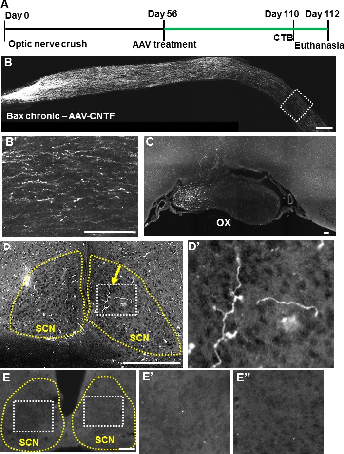
Visual target in the brain is reinnervated after postponed AAV-CNTF treatment in Bax−/− mice. (A) Time course of chronic experiment, with longer duration for regeneration (highlighted in green). (B) Sectioned optic nerve with CTB-traced RGC axons 16 weeks after injury and 8 weeks after intravitreal viral administration. High-magnification micrograph of distal region shown in (B′). (C–D) Coronal brain sections showing regenerating axons in the optic chiasm (C) and suprachiasmatic nucleus (SCN) (D), including at least one branched axon (yellow arrow) inside the visual target SCN (yellow outline). (D′) High magnification of the white boxed area in (D). (E) Representative coronal brain section of a control animal (i.e., Bax KO without AAV-CNTF) showing absence of regenerating axons. (E′, E′′) High magnification of the white boxed area in (E). OX, optic chiasm; SCN, suprachiasmatic nucleus. Scale bars: 150 μm.