
Figure 1.
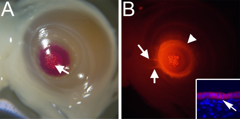
DiI labeling in embryonic chicken cornea. Brightfield (A) and immunofluorescent (B) images of the same DiI-labeled E10 cornea. Arrow in A shows crystals on CE surface. Arrowhead in B indicates sharp border of DiI diffusion (red); arrows in B show DiI-labeled nerves. Inset in B shows a section of DiI (red)-labeled cornea with nuclei stained with DAPI (blue); arrow in inset indicates Bowman's layer separating CE (above) from stroma (below).