Fig. 2.
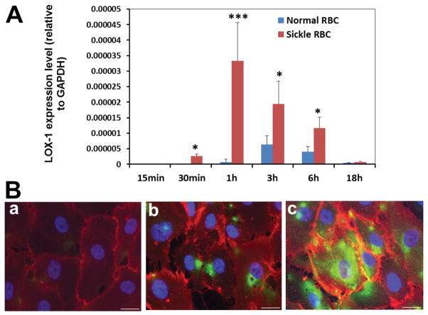
Sickle red blood cells induce LOX-1 gene expression in human brain microvascular endothelial cells (HBMVECs). A: HBMVECs were incubated with sickle RBC or normal RBC for 15 min–18 h. The expression of LOX-1 mRNA was assessed using real-time PCR. Data are presented as the means ± standard deviation (SD), differences between sickle RBC and normal RBC incubation groups at each time point were analyzed using Student’s t-test (***P value < 0.001, *P value < 0.05. n = 3 in each group; representative results of three independent experiments). Other experiments were carried out using heat-damaged normal RBCs. Heat damaging control RBCs did not increase expression of LOX-1 in HBMVEC over non-damaged RBCs (data not shown). B: Immunofluorescent staining of cultured HBMVECs for LOX-1 (green) and endothelium marker CD31 (red) ; (a) non-treated HBMVECs, (b) HBMVECs treated with normal RBC for 6 h, (c) HBMVECs treated with SCD RBC for 6 h (scale bar = 20 μm).

