Fig. 3.
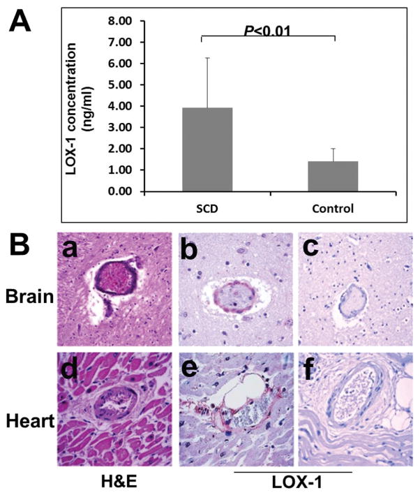
In vivo expression of LOX-1 in SCD patients. A: Measurement of circulating soluble LOX-1 (sLOX-1) concentrations by sandwich ELISA assay in SCD patient plasma and normal control plasma. Data are presented as means ± SD, difference between SCD and normal control was analyzed using Student’s t-test (n = 22 in SCD group and n = 9 in control group). B: Increased expression of LOX-1 in brain vascular endothelial cells of SCD patient. (a) H&E stain of midbrain autopsy tissue from a SCD patient showed thrombus formation and occlusion of the vessels, (b) IHC staining of the same SCD patient’s brain for LOX-1 showed increased expression of LOX-1 in vascular endothelial cells, (c) the expression of LOX-1 protein is almost undetectable by immunostaining in the control non-SCD brain tissue of a patient who died from trauma.

