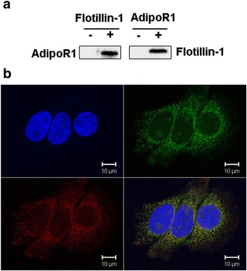
Fig. 5.

AdipoR1 protein interacted with Flotillin-1. The proteins that interacted with AdipoR1 were analyzed by co-immunoprecipitation (CO-IP) and confocal laser scanning microscopy (CLSM). MCF-7 cells were harvested after 48 h in culture, then total cellular proteins were extracted and CO-IP’d (co-immunoprecipitated) with anti-AdipoR1 antibodies followed by anti-Flotillin-1 antibodies (a). Then, the above proteins were then Co-IP’d with anti-Flotillin-1 antibodies followed by AdipoR1 antibodies (a). AdipoR1 is localized to the cell membrane of EC. After MCF-7 cells were co-transfection with plasmids pEGFP-C1-Flotillin-1 (green) and pDS-RED1-N1-AdipoR1 (red) for 48 h, the confocal scanning images demonstrated that the Flotillin-1 recombinant protein was localization to the cell membrane. The nuclei of the cells (blue) were stained by DAPI. The overlaid image indicates that AdipoR1 overlapped with Flotillin-1 at the MCF-7 cell membrane (b)
