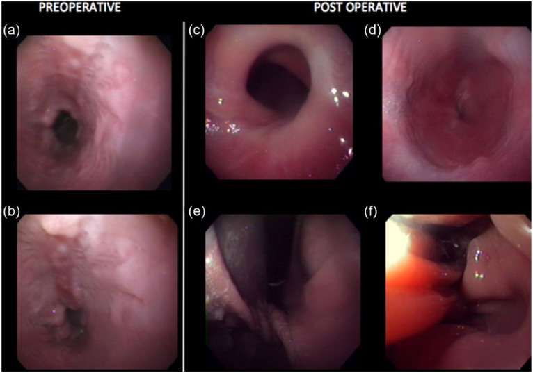
Figure 4.
Pre- and postsurgical endoscopy images. (a,b) Note the severe esophagitis with irregularly marginated, multifocal erosions. A significant amount of bilious reflux was noted. (c) Note the displaced and narrowed esophageal sphincter. (d) Note the mural twist at the cardia just caudal to the diaphragm. (e) Note the normal rugal folds of the fundus. (f) Note the structural abnormalities following the procedure, including the ‘tented’ margins of the lower esophageal wall

