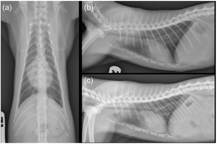
Figure 5.
Postoperative lateral and dorsoventral thoracic radiographs. (a) Ventrodorsal projection. Note the decreased size of the thoracic cavity compared with Figure 1 (a). This finding is likely due to resolved air trapping. Note also the resolution of the previously described moderate esophageal distention. (b) Right lateral projection. Note the lack of gas within the esophagus and the resolution of the previously described soft tissue mass at the level of the caudal esophagus. Mild, normal volume of mixed gas–fluid opacity is present within the fundus. Note also the normal contour of the ventral thoracic body wall with the more normally positioned sternebral segments due to resolved respiratory distress. (c) Left lateral. Note the poorly defined increase in soft tissue opacity just dorsal to the caudal vena cava and cranial to the diaphragm. Note the abnormal small volume of mixed fluid–gas opacity persistent within the fundus and the normal mixed gas–fluid opacity within the pylorus. Findings are consistent with the surgically induced malposition of the stomach

