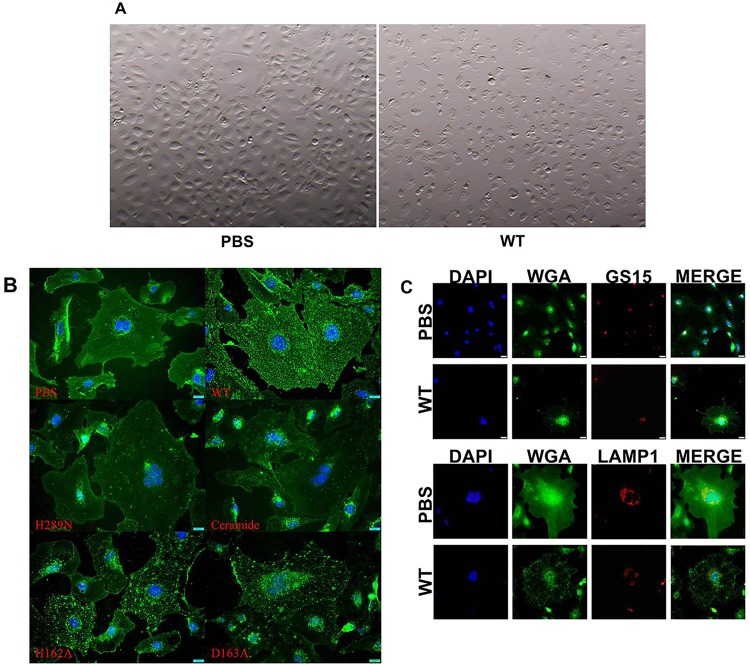
FIG 4 .
β-Toxin causes membrane alterations of HAECs. Images of HAECs under a dissecting microscope after treatment with β-toxin for 24 h are shown. (A) PBS-treated cells showing no change in cell morphology and retaining a cobblestone shape and HAECs treated with wild-type (WT) β-toxin, mutants, and ceramide-inducing cell rounding. (B) HAECs treated with β-toxin displaying granular distribution of wheat germ agglutinin (WGA) staining, PBS-treated cells show smooth even WGA staining throughout the cells, HAECs treated with wild-type or biofilm ligase mutants showing altered (speckled) cell membrane staining, treatment with the SMase mutant or ceramide appearing not to alter WGA cell membrane staining. (C) β-Toxin-induced speckling of HAECs resulting in the change to cell membrane morphology not due to the formation of endosomes from the Golgi apparatus or lysosomes. The top panel shows WGA does not colocalize with GS15 in β-toxin-treated cells differently from PBS-treated control HAECs. The bottom panel shows that LAMP-1 does not colocalize with WGA speckles formed from β-toxin treatment in HAECs.

