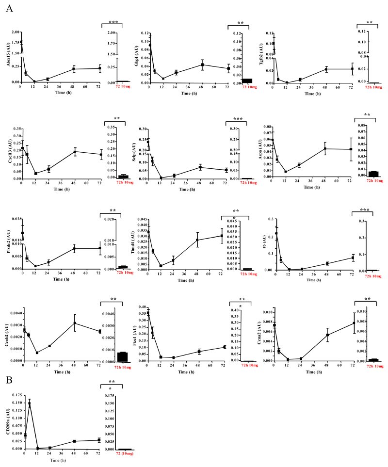
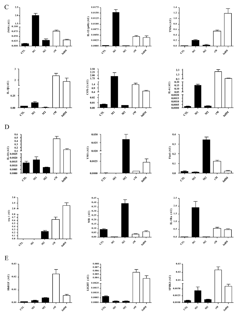
Figure 4. Validation of microarray analyses and phenotype of rM compared to in vitro-derived M1 and M2 macrophages.
Quantitative PCR for (A) genes most differentially expressed in rM versus pro-inflammatory macrophages validating original microarray findings including (B) CD209a, the monocytes-derive DC marker, which was included arising from the DC-like phenotype deduced in Figure 3. For comparisons to established M1/M2 cells, BMDMs were incubated with either LPS/INFγ (M1) and or IL-4 (M2) for 24h. RNA was extracted and probed for a range of typical (C) M1, (D) M2, and (E) M2b markers. Data are represented and analysed by ANOVA followed by Bonferroni multiple comparison tests. Values are expressed as means ± SEM of n=5-6 mice per group. ** P value < 0.01 and *** P value < 0.001.


