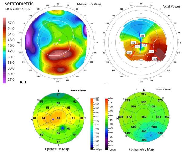
Figure 5.
An exceptional case of partial disagreement between maximum curvature (upper left), maximum keratometry (upper right), and minimum epithelial thickness (lower left). Note that the epithelium thinned focally over the maximum mean power (cone apex) but that a separate area of epithelial thinning at the superior periphery defined the map’s sector minimum. The area of pachymetric thinning is more inferotemporally displaced than the spot of maximum mean power (I = inferior; IN = inferonasal; IT = inferotemporal; N = nasal; S = superior; SN = superonasal; ST = superotemporal; T = temporal).

