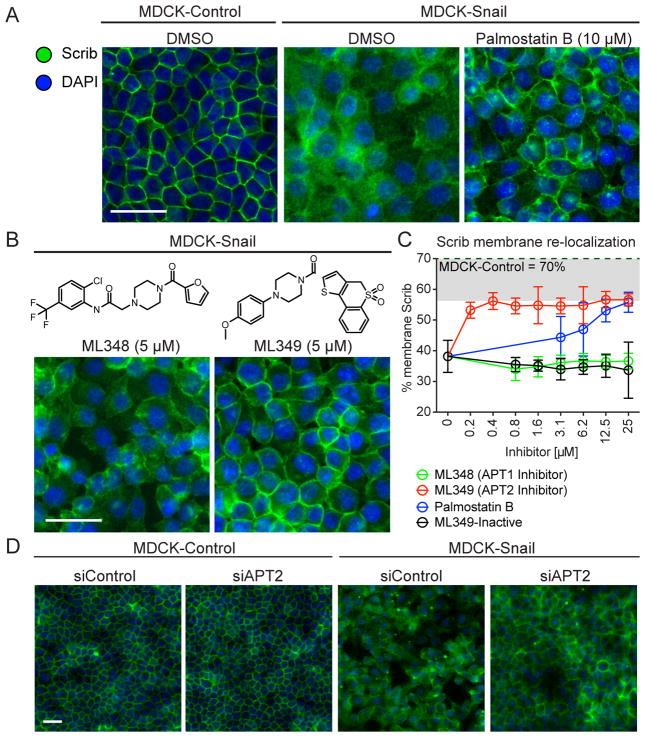
Figure 3. APT2 inhibition enhances Scrib perimeter localization in Snail-transformed cells.
(A–B) Representative immunofluorescence images of Scrib in MDCK-control and MDCK-Snail cells treated overnight with indicated inhibitors. (C) Dose-dependent enhancement of Scrib perimeter localization quantified by high-content automated microscopy, and processed as described in the Supporting Experimental Procedures. ML349-inactive lacks the two sulfone oxygens, which are required for APT2 inhibition in vitro. Standard deviations are shown for each condition. (D) APT2 knockdown in MDCK-Snail rescues Scrib membrane-localization. Representative images from high-content analysis of MDCK Control and Snail lines transfected with either non-targeting (siControl) or APT2 siRNA. Scale bar = 40 μm across each panel.

