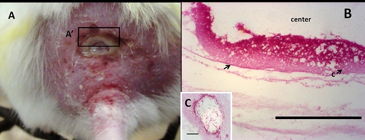
Fig 6. Gross morphology and ZN-stained section of the ulcer showing the distribution of acid-fast bacilli (i.e. M. ulcerans).
Panel A shows undermined ulcer at the back of a mouse. Panel B shows ZN stained thin section of A´ with an arrow showing bed of acid-fast bacilli localized at the edge of the ulcer. Panel C shows ring-like clustering unit of acid fast bacilli in panel B (the scale bar measures 10 μm).

