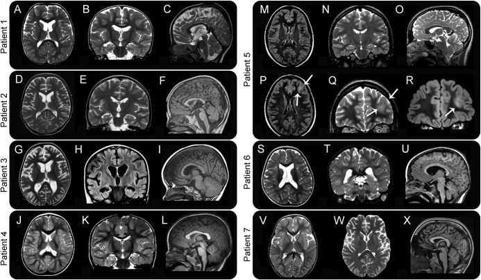
Figure 2. MRI characteristics of 7 patients with GNAO1 mutations.
For patients 1–6, a set of 3 images including an axial (A, D, G, J, M, and S), coronal (B, E, H, K, N, and T), and sagittal midline (C, F, I, L, O, and U) sequences is shown. For patient 5, in addition, 1 axial (P) and 2 coronal (Q and R) images are shown to illustrate better the neoplastic lesion in the left frontal lobe (arrows). For patient 7, axial images of 2 different investigations (V and W) and a sagittal image are shown to illustrate better the progression of atrophic changes. Images are at 1.5–3T. Images A–E, G, J, K, N, O, Q, S, T, and W are T2 weighted. Images F, I, L, U, and X are T1 weighted. Images H, M, P, and R are acquired as fluid attenuation inversion recovery (FLAIR) sequences. Patient 1: axial and coronal images show mild ventricular enlargement in the frontal horns, with concomitant hypoplasia of the caudate nuclei. The sagittal section shows a thin corpus callosum. Patient 2: axial and coronal images show mild ventricular enlargement in the frontal horns, with mild hypoplasia of the caudate nuclei, likewise seen in patient 1. The sagittal section shows a thin corpus callosum and a hypoplastic inferior vermis. Patient 3: axial and coronal images show moderate cortical atrophy, with ventricular enlargement and small caudate nuclei. The sagittal image shows a dysmorphic corpus callosum. Patient 4: no structural abnormality is visible on MRI. Patient 5: MRI shows an isolated abnormality (arrows) involving the anterolateral aspect of the left frontal lobe, including the cortex and underlying white matter, with increased signal intensity on both T2 and FLAIR images. Histopathology, after surgical removal of the lesion, showed characteristics consistent with a diffuse astrocytoma (WHO grade 2). Patient 6: axial and coronal images show dilated lateral ventricles; the sagittal cut shows a thin corpus callosum. Patient 7: the first axial MRI at age 2 (V) is normal, but a follow-up study at age 16 (W) shows loss of brain volume in generalized distribution. No abnormalities in the brainstem and cerebellum are visible in the sagittal image at age 16 (X).

