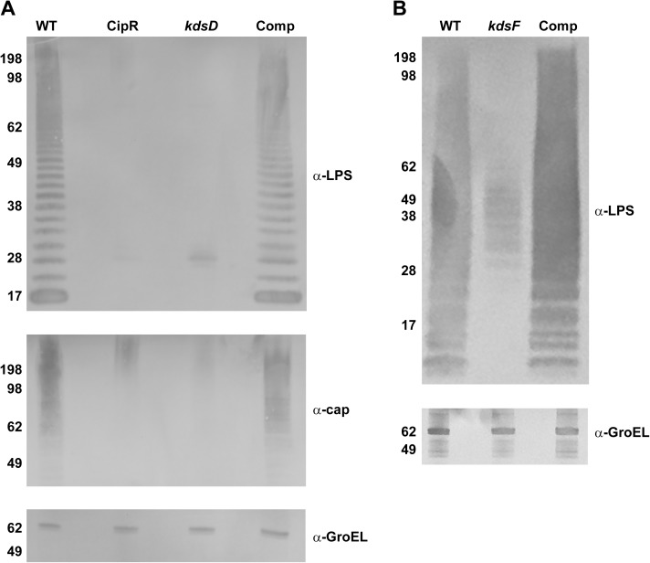
Fig 2. Western blot analysis of Francisella strains.
Pellets of the (A) F. tularensis strains: Schu S4 (WT), CipR, kdsD::ltrBL1, and kdsD complement (Comp) or (B) F. novicida strains: U112 (WT), kpsF::T20, and kpsF complement (Comp) were lysed. Extracts were run on SDS-PAGE gels at equal concentrations and blotted with various antibodies as indicated: monoclonal antibody to the O-antigen of LPS of F. tularensis or F. novicida; monoclonal antibody to the O-antigen of the F. tularensis capsule; or a polyclonal antibody to GroEL of both F. tularensis and F. novicida. Molecular masses are indicated on the left in KDa. A) The LPS and capsule profiles of the CipR and kdsD::ltrBL1 mutants were defective in comparison to the WT strain. However, these profiles were restored for the kdsD mutant when complemented with a functional gene on a plasmid. Equal loading of sample material was demonstrated when blotting the extracts with an antibody directed against the GroEL protein. B) The LPS profile of the kpsF::T20 mutant was defective in comparison to the U112 parent strain. However, the profile was restored for the kpsF::T20 mutant when complemented with a functional kdsD from F. tularensis was provided on a plasmid. Equal loading of sample material was demonstrated when blotting the extracts with an antibody directed against the GroEL protein.

