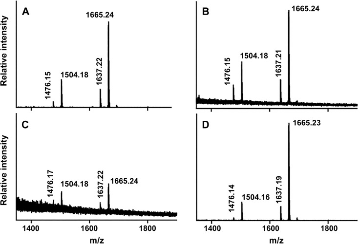
Fig 3. Characterization of the F. tularensis lipid A structure by MALDI-TOF mass spectrometry.
LPS extracts from wild type Schu S4 (A), CipR (B), kdsD::ltrBL1, (C) and complemented kdsD mutant were analyzed by negative ion mode MALDI-TOF mass spectrometry. Monoisotopic mass/charge values of the four most prominent species within each spectrum are reported; these values correspond with the expected molecular weights of F. tularensis lipid A and its known variants as previously reported [29].

