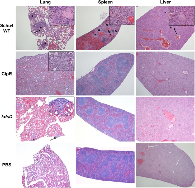
Fig 8. Pathology of mice challenged intranasally with F. tularensis 5 days post-challenge.
Mice were challenged intranasally with Schu S4 WT (131 CFU), CipR mutant (1,750 CFU), or kdsD::ltrBL1 (6,000 CFU). Also included as a control for these studies were mice receiving PBS alone by intranasal administration. The strains used for challenge and HE stained organ (lung, spleen, and liver) are as indicated. All samples shown are at 5 days post-challenge when the Schu S4 challenged mice were moribund; in contrast, the mutant challenged mice displayed no clinical signs of infection at this time point. Schu S4 WT, Lung (4x)–multifocal areas of inflammation and necrosis (*); arrow indicates the inset area. Inset (40x)–necrosis admixed with inflammatory cells. Spleen (4x)–coalescing areas of necrosis (*) affecting red pulp and white pulp; arrow indicates the inset area. Inset (40x)–necrosis admixed with inflammatory cells. Liver (4x)–Single focus of necrosis (*); arrow indicates the inset area. Insert (40x)–necrosis with few inflammatory cells. CipR mutant, Lung (4x)–diffuse coalescing necrotic areas. Inset (40x)–necrosis admixed with inflammatory cells. Spleen (4x)–normal. Liver (4x)–normal. kdsD::ltrBL1, Lung (4x)–few foci of necrosis (*); arrow indicates the inset area. Inset (40x)–necrosis admixed with inflammatory cells extends to the surface of the lung. Spleen (4x)–normal. Liver (4x)–normal.

