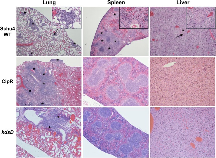
Fig 10. Pathology of mice challenged by small particle aerosol with F. tularensis.
The strains used for aerosol challenge (Schu S4 WT, CipR, and kdsD::ltrBL1) and the HE stained organ (lung, spleen, and liver) are as indicated. The mice challenged with Schu S4 were collected at Day 5 when all mice had succumbed to infection. The mice challenged with the CipR or kdsD::ltrBL1 mutant survived till the end of the study (Day 21) and displayed no clinical signs of infection. Schu S4 WT: Lung (4x)–multifocal areas of inflammation and necrosis (*); arrow indicates the inset area. Inset (40x)–necrosis admixed with inflammatory cells. Spleen (4x)–diffuse coalescing areas of necrosis (*) affecting red pulp and white pulp. Inset (40x)–necrosis (*) admixed with inflammatory cells; arrow indicates the inset area. Liver (10x)–Single focus of necrosis (*); arrow indicates the inset area. Insert (40x)–necrosis with few inflammatory cells. CipR: Lung (4x)–Diffuse coalescing areas of necrosis (*) with inflammatory cells that extend to the surface of the lung. Spleen (4x)–normal. Liver (10x)–normal. kdsD::ltrBL1: Lung (4x)–foci of necrosis (*) centered on a large airway. Spleen (4x)–normal. Liver (10x)–normal.

