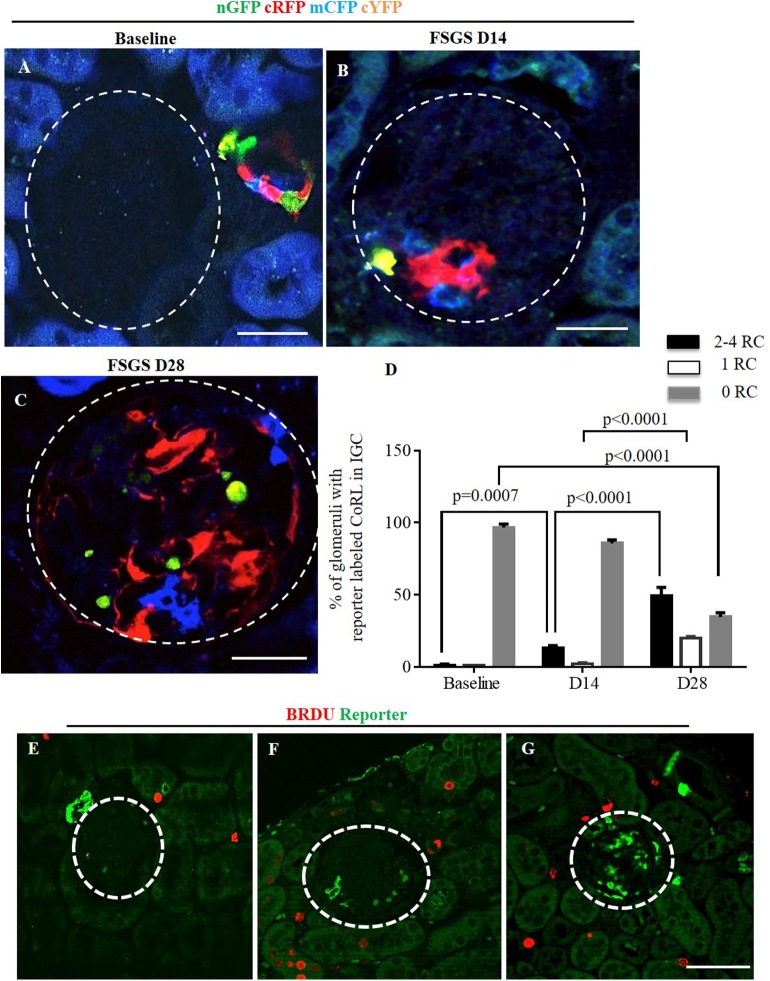
Fig 3. Multi-colored reporters of CoRL are detected in glomerular tufts of Ren1cCre /R26R-ConfettiTG/WT mice with FSGS.
Confocal images showing four CoRL reporter colors detected without the use of antibodies–nGFP (green), cRFP (red), mCFP (blue) and cYFP (yellow). (A) All four reporters are restricted to the JGC at baseline, and are not detected in the glomerular tuft (dashed white circles). At D14 FSGS (B) and at D28 FSGS (C), all four CoRL reporter colors were detected in a subpopulation of cells in the glomerular tufts. (D) Graph showing that the percentage of glomeruli with reporter positive CoRL within the tuft was higher at D28 of FSGS and that these glomeruli contained 2–4 clones. (E) Represenative image showing all four reporters (converted to green color) and BRDU(red) do not co-localize at baseline. BRDU is present in some tubular epithelial cells as expected, but is not readily detected in JGC or the glomerular tuft. (F) BRDU staining increased at D14 of FSGS, but BRDU positive cells are not present in the JGC and glomerular tuft. (G) At D28 of FSGS there is an increase in the number of reporter labeled cells present on the glomerular tuft, however there is no overlap of reporters with BRDU.

