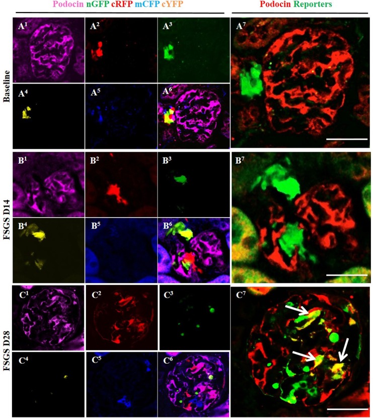
Fig 4. Labeled cells of renin lineage (CoRL) co-express podocin in glomeruli of Ren1cCre /R26R-ConfettiTG/WT mice with experimental FSGS.
(A1-A7) At baseline: (A1) Confocal image shows podocin antibody staining (magenta). (A2-A5) All 4 CoRL reporters (green, red, blue, yellow) can be detected without the use of antibody. (A6) Composite image of all 4 reporters and podocin staining. (A7) For ease of viewing, all 4 confetti reporter channels have been converted to green, and podocin has been converted to red, so that co-localization can be visualized as yellow. All four confetti reporters are seen in the JGC, with no overlap with podocin staining. (B1-B7) At day 14 FSGS: (B1) There is a segmental decrease in podocin staining in the left lower quadrant of the glomerular tuft. (B2-B6) Multi-clonal CoRL (red, yellow and green) are detected in glomerular tuft, but do not co-localize with podocin. (B7) The CoRL reporters in the tuft do not co-localize with podocin staining. (C1-C7) At D28 FSGS: (C1-C5) Podocin and multi-clonal CoRL (red, yellow and green) are detected in the glomerular tuft. (C6) Composite image of all four reporters and podocin staining. (C7) CoRL reporters co-localize with podocin, creating a yellow color (arrows indicate examples).

