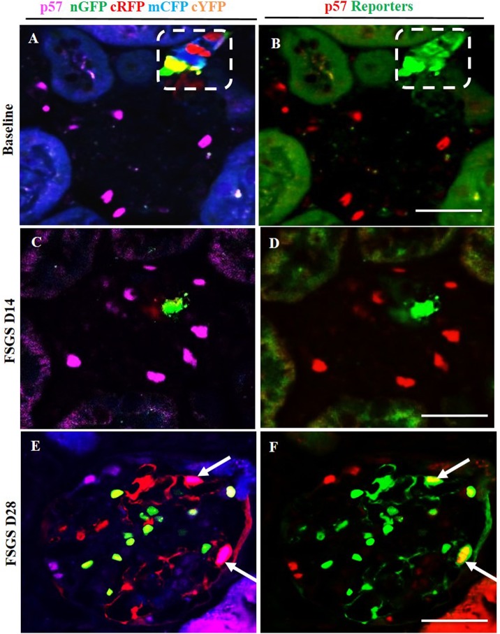
Fig 6. Labeled cells of renin lineage (CoRL) co-express p57 in glomeruli of Ren1cCre /R26R-ConfettiTG/WT mice with experimental FSGS.
The confocal images in the left column (A-C) represent p57 staining (nuclear, magenta) detected by antibody, and 4 CoRL reporters (green, red, blue, yellow) detected without antibody. The confocal images in the right column represents the same image on the left, but for ease of viewing, all 4 confetti reporter channels have been converted to green, and p57 has been converted to red, so that co-localization can be visualized as yellow. (A, B) At Baseline: all four CoRL reporter colors are restricted to the JGC (dashed white box), and p57 staining is restricted to the glomerular tuft. (B) all four Confetti CoRL reporters (green) are seen in the JGC, with no overlap with p57 (red). (C, D) At D14 FSGS: (C) There is a segmental decrease in p57 staining in the right upper quadrant of the glomerular tuft. (D) The CoRL reporters in the tuft do not co-localize with p57 staining. (E, F) At D28 FSGS: (E) Multi-clonal CoRL are detected in the glomerular tuft. (F) CoRL reporters co-localize with p57, creating a yellow color (arrows indicate examples).

