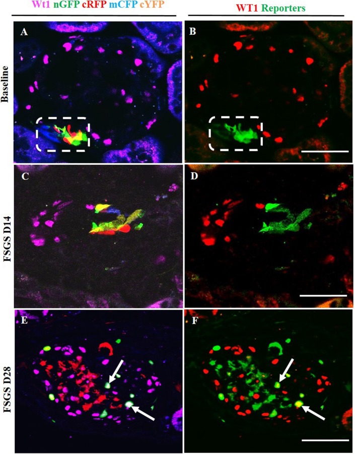
Fig 7. Labeled cells of renin lineage (CoRL) co-express WT-1 in glomeruli of Ren1cCre /R26R-ConfettiTG/WT mice with experimental FSGS.
The confocal images in the left column (A-C) represent WT-1 staining (nuclear, magenta) detected by antibody, and 4 CoRL reporters (green, red, blue, yellow) detected without antibody. The confocal images in the right column represents the same image on the left, but for ease of viewing, all 4 confetti reporter channels have been converted to green, and WT-1 has been converted to red, so that co-localization is visualized as yellow. (A, B) At Baseline: (A) All four CoRL reporter colors are restricted to the JGC (dashed white box), and WT-1 staining is restricted to the glomerular tuft. (B) All four Confetti CoRL reporters (green) are seen in the JGC, with no overlap with WT-1 (red). (C, D) At day 14 FSGS: (C) There is a segmental decrease in WT-1 staining in the lower half of the glomerular tuft. Multi-clonal CoRL are detected in glomerular tuft, but do not co-localize with WT-1. (D) The CoRL reporters in the tuft do not co-localize with WT-1 staining. (E, F) At D28 FSGS: (E) Multi-clonal CoRL are detected in the glomerular tuft. (F) CoRL reporters merge with WT-1, creating a yellow color (arrows indicate examples).

