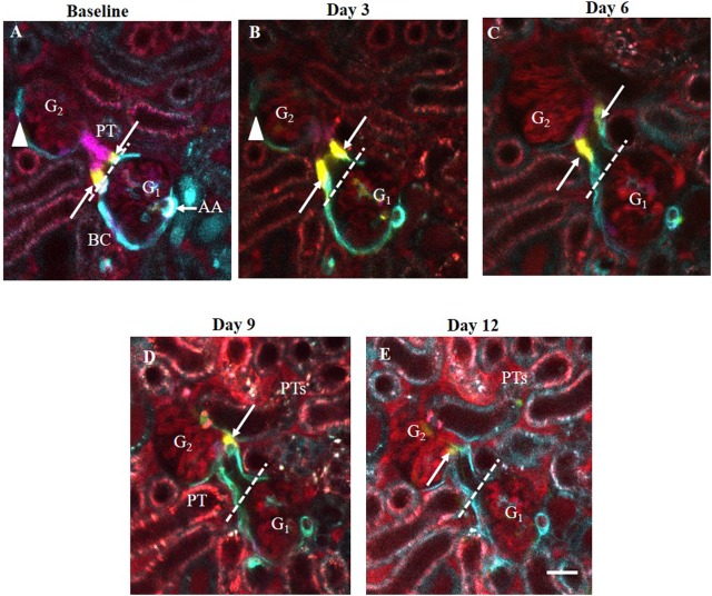
Fig 10. Serial intravital MPM imaging of the same glomeruli (G1-G2) in the same Ren1d-Confetti mouse kidney over two weeks after IgG-induced podocyte injury.
(A) At baseline, several multi-color CoRL form the terminal portion of afferent arteriole (AA, mostly expressing blue, membrane-targeted CFP), the parietal Bowman’s capsule and the early proximal tubule (PT). Bar is 20 μm. Note the 1–2 yellow-labeled (cytosolic YFP expressing) CoRL (arrows) localized exactly at the glomerulo-tubular junction (dashed line). (B-E) Three-to-twelve days after IgG injection, the same YFP+ cells in the same glomerulus (G1) moved continuously away from the glomerulo-tubular junction and deeper into the proximal tubule. During the same time, CFP+ cells along the parietal Bowman's capsule of the other glomerulus (G2) disappeared from Bowman's capsule (arrowhead) between D3-6 (B-C). Plasma was labeled red using Alexa594-albumin. Note the significantly increased albumin content in G and PT fluid and within PT cells in Day 9–12 (intense red).

