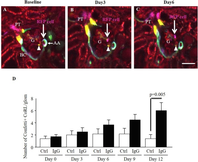
Fig 11. Tracking the migration of individually labeled single CoRL cells in the same Ren1d-Confetti mouse kidney after IgG-induced podocyte injury using serial intravital MPM imaging.
(A) At baseline, several multi-color CoRL surround the terminal portion of the afferent arteriole (AA), line the parietal Bowman’s capsule and the early proximal tubule (PT). Bar is 20 μm. Note the magenta-labeled (cytosolic RFP expressing) single CoRL at the terminal AA closest to the glomerular tuft (RFP cell, arrow). (B) Three days after IgG injection, what is likely the same RFP+ cell in the same glomerulus (G) appears detached from the AA and localizes in the glomerular tuft (arrow). (C) Six days after IgG injection the RFP+ cell appears localized around a glomerular capillary (arrow). In contrast to the migrating RFP+ cell, a YFP+ CoRL (yellow, cytosolic YFP expressing) appears stationary in the intraglomerular mesangium (arrowhead). Plasma was labeled red using Alexa594-albumin. (D) Analysis of the total number of Confetti+ CoRL in the glomerular tuft in time-control (Ctrl, n = 5) and IgG-injected (IgG, n = 4) mice during the first two weeks of podocyte injury.

