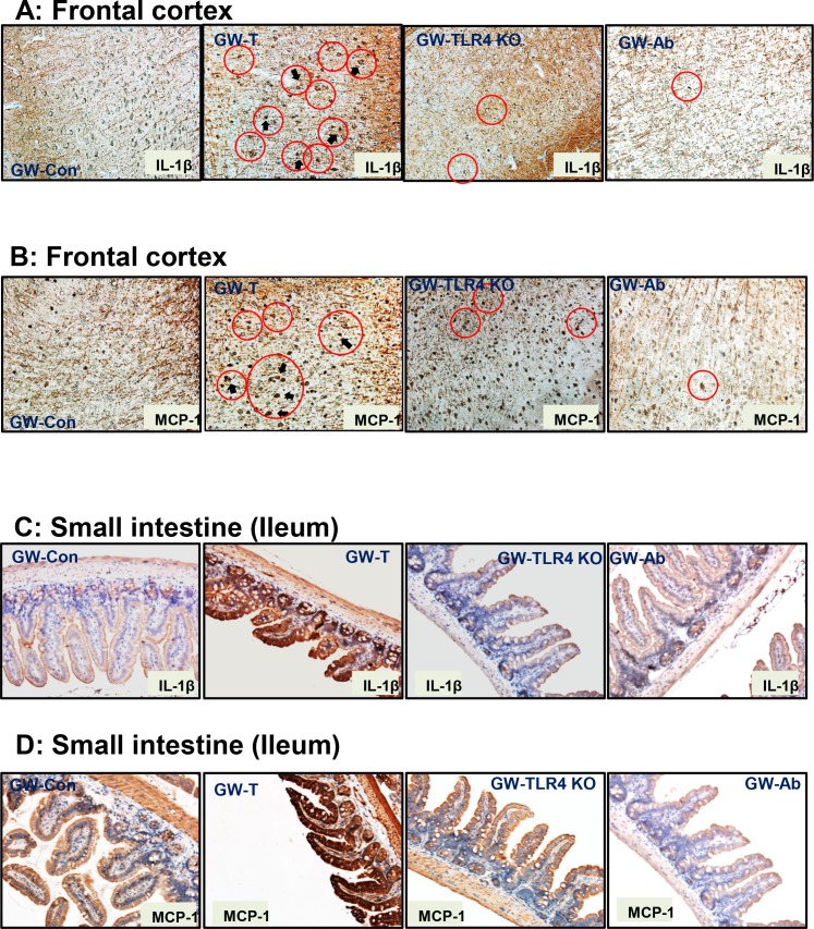
Fig 9. Gulf war chemical exposure-induced changes in gut microbiome modulates neuroinflammation and intestinal cytokine release.
A. Frontal cortex tissue slices were probed for IL-1β immunoreactivity in control (GW-Con, treated (GW-T), TLR4 knockout mice treated with GW Chemical exposure (GW-TLR4KO) and antibiotic treated (GW-Ab) groups. Specific immunoreactivity to IL-1β as evident by dark brown spots are indicated by black arrows and areas of interest are outlined by red circles. B. Frontal cortex tissue slices were probed for MCP-1 immunoreactivity in control (GW-Con, treated (GW-T), TLR4 knockout mice treated with GW Chemical exposure (GW-TLR4KO) and antibiotic treated (GW-Ab) groups. Specific immunoreactivity to MCP-1 as evident by dark brown stain/spots are indicated by black arrows and areas of interest are outlined by red circles. C. Small intestine tissue slices were probed for IL-1β immunoreactivity in control (GW-Con, treated (GW-T), TLR4 knockout mice treated with GW Chemical exposure (GW-TLR4KO) and antibiotic treated (GW-Ab) groups. D. Small intestine tissue slices were probed for MCP-1 immunoreactivity in control (GW-Con, treated (GW-T), TLR4 knockout mice treated with GW Chemical exposure (GW-TLR4KO) and antibiotic treated (GW-Ab) groups.

