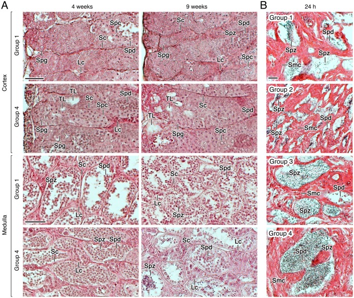
Fig 4. Testicular development of males treated with rFsh and rLh in experiment 1.
(A) Representative photomicrographs of histological sections from the cortical and medullar regions of the testis of Group 1 and 4 stained with hematoxylin and eosin after 4 and 9 weeks of treatment. (B) Photomicrographs of histological sections from the testicular sperm duct stained with hematoxylin and eosin from Groups 1 to 4 at 24 h after rLh injection. Scale bars, 20 μm. Sc, Sertoli cell; Lc, Leydig cell; Spg, spermatogonia; Spc, spermatocyte; Spd, spermatid; Spz, spermatozoa; TL, forming tubular lumen.

