
Fig. 4.
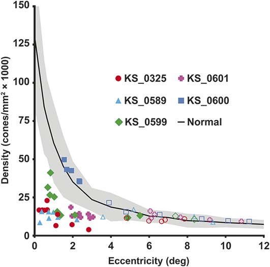
Cone photoreceptor density inside and outside lesions. Density was sampled in all subjects within (filled symbols) and outside their BVMD lesion (open symbols). Cone density is significantly reduced within the lesions, but returns to normal outside lesions. Normative data30 are shown as mean (solid line) ± two SD (shaded region).