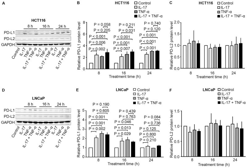
Fig. 2.
Effects of IL-17 and/or TNF-α on PD-L1 and PD-L2 protein expression in human cancer cell lines HCT116 and LNCaP. (A to C) HCT116 cells and (D to F) LNCaP cells were treated with IL-17 (20 ng/ml), TNF-α (10 ng/ml), or a combination of both for the indicated time. PD-L1 and PD-L2 protein expression was analyzed using Western blot analysis. GAPDH was probed for protein loading control (A and D). The relative protein levels were presented as the ratio of PD-L1/GAPDH or PD-L2/GAPDH (B–C and E–F), which was normalized against the ratio of the control group (arbitrarily designated as “1”). Data were represented as means ± SD of three independent experiments.

