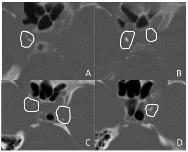
Figure 1. Representative Computed Tomography Images.
Images are shown in bone windows from two patients with intracranial internal carotid artery (ICA) calcifications.
Panels A and B. Two contiguous axial images from Patient 1 in panels A and B show manually drawn regions of interest surrounding ICA calcification, with care taken to avoid including adjacent skull structures. This patient had Modified Woodcock Visual Scale (MWVS) scores of 2 and 3 for the right ICA (A) and 0 and 1 for the left ICA (B). The total Agatston-Janowitz (AJ) 130 score was 334 for the right ICA and 23 for the left. This patient had a right-sided acute anterior circulation infarction.
Panels C and D. Two contiguous axial images from Patient 2 in panels C and D show similarly drawn regions of interest surrounding ICA calcifications. This patient had MWVS scores of 1 and 0 for the right ICA (C) and 2 and 3 for the left ICA (D). The total AJ-130 score was 84 for the right ICA and 198 for the left. This patient had a left-sided acute anterior circulation infarction.

