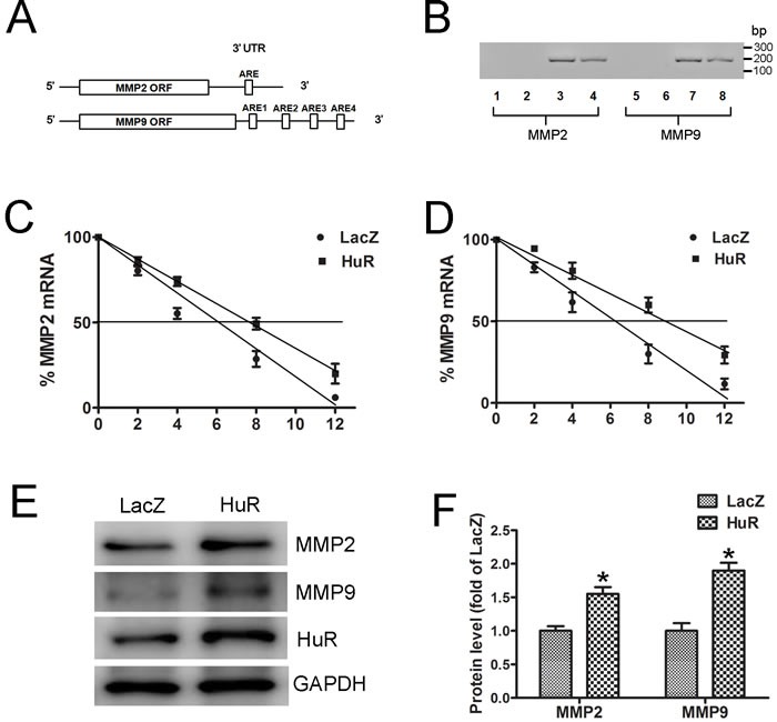
Figure 2. MMP2 and MMP9 are targets of HuR.

A. Schematic representations of predicted AREs in the 3’ UTR of MMP2 and MMP9 mRNAs are depicted. B. RNA immunoprecipitation was carried out with the anti-HuR antibody or control IgG. Lanes 1 and 5, no template PCR control; lanes 2 and 6, IgG RNA immunoprecipitation; lanes 3 and 7, Anti-HuR RNA immunoprecipitation; and lanes 4 and 8, 10% input. C. and D. MOVAS cells were transfected with LacZ or HuR plasmid for 24 hours and then treated with actinomycin D (5 μg/mL). The levels of MMP2 (C) and MMP9 (D) mRNAs were determined by real-time PCR (n = 4). E. Western blot analysis to detect MMP2 and MMP9 expression in MOVAS cells after LacZ or HuR transfection. F. Quantitative analysis of MMP2 and MMP9 protein levels in SMCs (n = 4). *p < 0.05 vs LacZ.
