
Figure 2. KLK6 activates proMMP2.
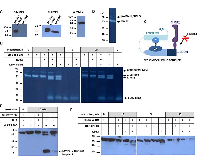
(A) Western blot analysis of SH-SY5Y cell supernatants for detection of MMP9, TIMP2, and MMP2. Positive controls for MMP9 included U2OS cells stimulated with TPA and for TIMP2 MDA-MB-231 cells. (B) Gelatin zymography showed two bands that correspond, to proMMP2 and potentially to proMMP2/TIMP2, respectively. (C) Schematic diagram of the proMMP2/TIMP2 complex showing coverage of the C-terminal domain of proMMP2 that is recognized by the anti-MMP2 antibody that could account for the band detected around 100 kDa. (D) Time course of proMMP2 activation in SH-SY5Y cell supernatant. EDTA did not inhibit proMMP2 activation. Proteolytic activities were detected by gelatin zymography. (E) Activation of proMMP2 monitored by Western blotting with a specific antibody for the C-terminus of proMMP2. (F) Time-course of proMMP2 activation indicated by the gradual decrease of the proMMP2 form. The first antibody used binds specifically the propeptide of proMMP2, which is removed upon activation. The experiments were carried out three times.