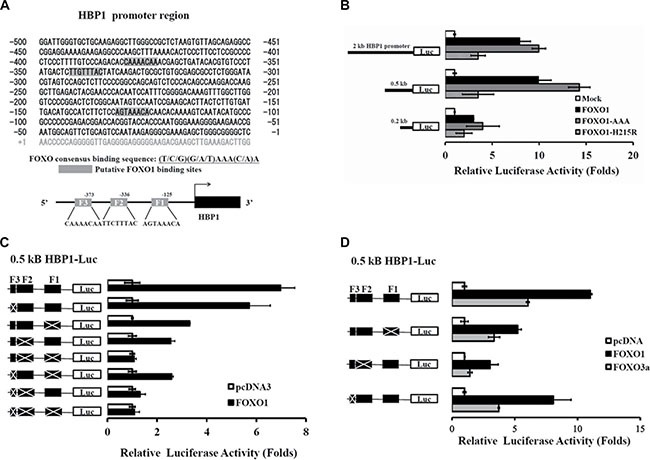
Figure 4. Identification of FOXO1 response elements in the HBP1 promoter.

(A) Schematic diagram of the HBP1 proximal promoter containing three potential FOXO1 binding sites at positions –132 to –125, –343 to –336, and –380 to –373 bp (depicted as F1, F2, and F3, respectively) from the transcriptional start site as predicted by MAPPER Search Engine. (B) Relative activation of FOXO1 and its mutants on various lengths of the native HBP1 promoters. HEK-293T cells were co-transfected with luciferase reporters fused with the indicated lengths of the HBP1 promoter as well as the expression plasmid of wild-type FOXO1 or FOXO1 mutant (FOXO1-AAA or FOXO1-H215R). Luciferase activity normalized to β-galactosidase was determined 24 h after transfection and represented as means ± S.E.M. from three separate experiments. (C–D) A 0.5-kb HBP1 promoter-luciferase construct with a series deletion combination of F1, F2 and/or F3 was co-transfected with a (C) FOXO1 or (D) FOXO1 or FOXO3a expression plasmid into HEK-293T cells. After 24 h of incubation, luciferase activity relative to control (empty vector) was determined after normalization to β-galactosidase.
