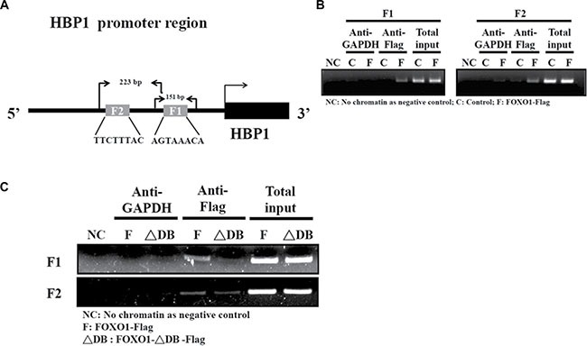
Figure 5. FOXO1 occupies its consensus binding sites in the endogenous HBP1 promoter.

(A) A schematic diagram indicates two primer sets designed for the PCR detection and the expected sizes of the PCR products in the chromatin immunoprecipitation (ChIP) assay. (B–C) ChIP assay was performed to determine the formation of FOXO1-HBP1 promoter complex in (B) HEK-293T and (C) HSC-3 cells. Cells were transfected with a pcDNA3 (Control; C), pcDNA3-FOXO1-Flag (FOXO1-Flag; F), or pcDNA3-FOXO1-ΔDB-Flag (FOXO1-ΔDB-Flag; ΔDB) plasmid for 48 h, followed by sequential fixation, immunoprecipitation with anti-Flag or GAPDH antibody, and PCR analysis with the primer sets indicated in (A).
