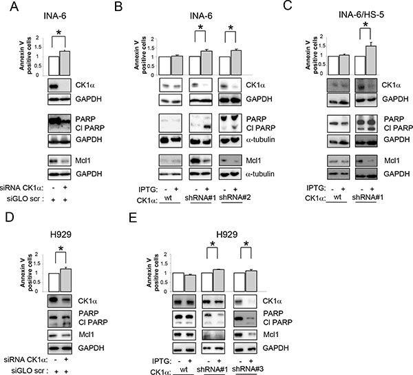
Figure 3. Effects of CK1 inhibition on MM cell survival.

Quantification of apoptosis through Annexin V/PI staining and FACS analysis (upper panel) and determination of PARP cleavage and Mcl1 protein expression (lower panel) in INA-6 (A–C) or H929 (D, E) silenced for CK1α. CK1α silencing was obtained with electroporation of ds oligonucleotides directed against CK1α (A, D) or using IPTG inducible CK1α shRNA cellular clones (shRNA#1, shRNA#2, shRNA#3, B,C,E). Cells were treated with IPTG 500 μM (INA-6, and H929 shRNA#1) or 1000 μM (H929 shRNA#3) for 7 days (INA-6 shRNA#1, and H929 shRNA#1 and 2) or 12 days (INA-6 shRNA#2). For the BM microenvironment model (C) INA-6 wt and shRNA#1 cells were treated with IPTG 500 μM for 6 days and subsequently plated on HS-5 stromal cells for an additional 4 days in the continuous presence of IPTG. In all experiments, IPTG was added also to wt INA-6 cells and H929 cells as control. GAPDH or α-tubulin was used as loading control. * indicates p < 0.05. Data represent the mean ± SEM of three independent experiments and are presented as arbitrary values over untreated cells.
