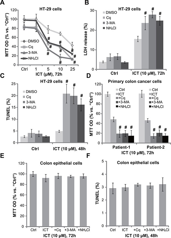
Figure 2. Autophagy inhibitors potentate icaritin-induced CRC cell death and apoptosis.

HT-29 cells (A–C), primary colon cancer cells (two lines, “Patient-1/−2”) (D), or the primary colon epithelial cells (E and F), pre-treated for 30 min with applied autophagy inhibitors: 3-methyladenine (3-MA, 1.0 mM), chloroquine (Cq, 5 μM) or ammonium chloride (NH4Cl, 2.5 mM), were subsequently treated with ICT (10 μM) for designated time; Cell viability, cell death and apoptosis were tested by MTT assay (A, D and E), LDH release assay (B) and TUNEL staining assay (C and F), respectively. Experiments in this figure were repeated three times, and similar results were obtained. “DMSO” stands for 0.1% DMSO of vehicle control. Data were expressed as mean ± SD, experiments were repeated five times. n = 5 for each assay. #p < 0.05 vs. “DMSO” group.
