
Figure 5. Expression of the candidate genes in highly and minimally invasive/migratory subclones.
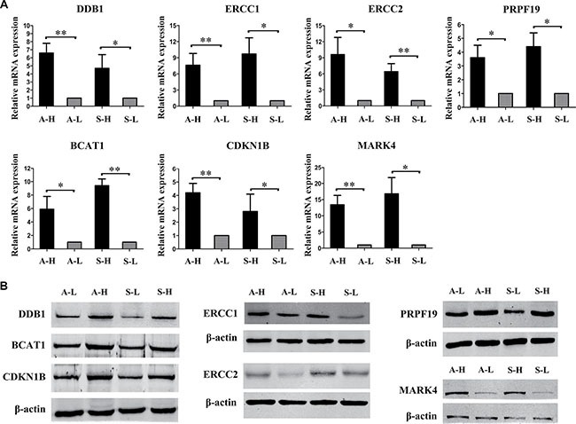
(A) RT-PCR and (B) Western blot analysis consistently confirmed that DDB1, ERCC1, ERCC2, PRPF19, BCAT1, CDKN1B and MARK4 expression were all greater in A-H/S-H cells than in A-L/S-L cells. Error bars represent the SEM, n = 3 (*P < 0.05. **P < 0.01).