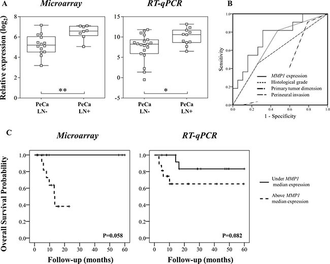
Figure 3.

(A) Microarray and RT-qPCR data revealed higher MMP1 expression in primary tumors from patients that presented inguinal lymph node metastasis (LN+). (B) MMP1 was a better predictor of lymph node status compared with histological grade, primary tumor size (T1-T4) and perineural invasion. Area under the curve (AUC) for MMP1 expression: 0.751, histological grade: 0.672; primary tumor size (T1-T4): 0.376 and perineural invasion: 0.596. (C) Overall survival analyses of PeCa patients according to MMP1 expression patterns detected by microarray and RT-qPCR analyses. Kaplan-Meier curves show high expression of MMP1 (defined as values above the median expression) associated with shorter survival.
