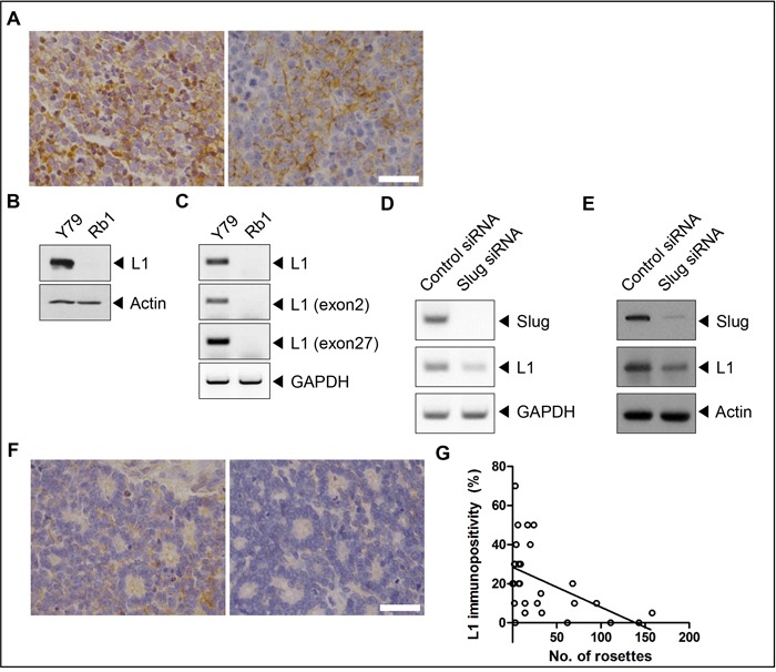
Figure 1. L1 is differentially expressed in retinoblastoma.

A. L1-positive retinoblastoma cells in areas with compactly packed tumor cells in retinoblastoma tissues. B and C. The expression of L1 in Y79 and SNUOT-Rb1 cells by (B) Western blot analysis and (C) RT-PCR. D and E. The expression of L1 in control and Slug-depleted Y79 cells by (D) Western blot analysis and (E) RT-PCR. F. No to weak immunopositivity of L1 in areas with multiple Flexner-Wintersteiner rosettes in retinoblastoma tissues. G. The correlation between the number of Flexner-Wintersteiner rosettes and the proportion of L1-positive cells in each tumor sample. Rb1, SNUOT-Rb1 cells. Scale bar, 25 μm.
