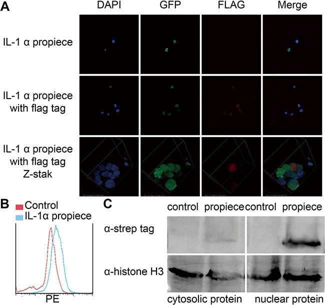
Figure 2. IL-1α propiece is located in nuclei.

A. Jurkat-proIL-1α with FLAG tag (red) was analyzed by immunofluorescence and confocal microscopy (40X). Nuclear DNA was visualized by DAPI staining (blue). B. The nuclei of the Jurkat-proIL-1α was analyzed by flow cytometry. C. Jurkat-proIL-1α were separated for cytosolic and nuclear proteins, which were then blotted for IL-1α propiece with anti-strep antibodies. Data shown are the representatives of at least three independent experiments.
