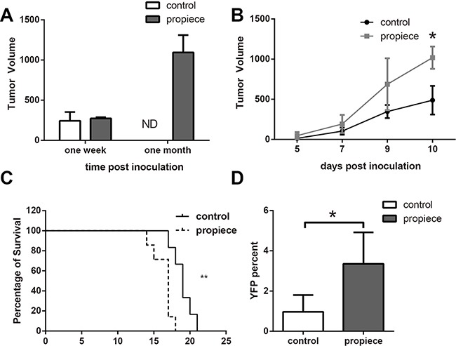
Figure 4. IL-1α propiece promotes the in vivo development of T-ALL.

A. Five million Jurkat cells in 100 μl of BD Matrigel were inoculated into nude mice subcutaneously. Tumor sizes were measured at one week and one month after inoculation. B. Murine p388 cells (105) were inoculated subcutaneously into DBA mice. Tumor sizes were measured every other day in a blinded fashion. C. DBA mice inoculated with p388 cells subcutaneously developed metastatic leukemia. The survival curves of the mice that received p388 cells expressing IL-1α propiece or the control vector were plotted. Statistical differences in survival times were determined using Kaplan–Meier survival curves and X 2 analysis. D. The YFP positive cells in peripheral blood of the p388 tumor-bearing mice were analyzed by flow cytometry. The experiments were performed with five mice per group. Data shown are the representatives of at least three independent experiments. Results are expressed as mean ± SD. * p<0.05, **p<0.01.
