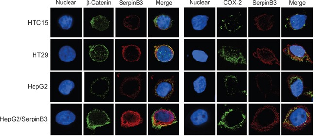
Figure 4. Immunofluorescence analysis of SerpinB3, COX-2 and β-Catenin.

Immunofluorescence staining of HTC15, HT29, HepG2 and HepG2/SerpinB3 cell lines after 48 hour of culture. Left panel: Double staining for SerpinB3 and β-Catenin proteins. Right panel: Double staining for SerpinB3 and COX-2 proteins. Proteins were visualized under a fluorescent microscope: TRIC (red, SerpinB3), FITC (green, β-Catenin and COX-2) and DAPI (blue, nuclei). Original magnification 400 X
