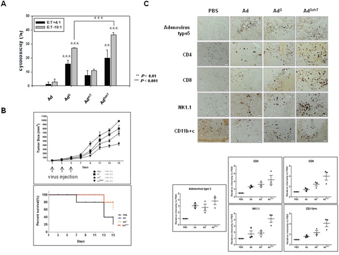
Figure 4. The anti-tumor effect of adenoviruses expressing mGM-CSF and shmTGF-β2.

The anti-tumor effect of oncolytic Ad-GMCSF, Ad-shTGFβ2 or Ad-GMCSF-shTGFβ2 was confirmed by ex vivo (A) and in vivo (B) experiments. A. B16BL6-CAR/E1B55 cells infected with each virus at an MOI of 50 were incubated with splenocytes isolated from 3 mice of each group of C57BL/6 for 4 h. The splenocyte cytotoxic activity was measured by an LDH assay. E:T means the ratio of total number of effector cells (splenocytes) to target cells (infected B16BL6-CAR/E1B55 cells). B. C57BL/6 tumor-bearing mice were treated with intratumoral injections of 1 × 10⁹ PFU/50 μL of adenoviruses on days 1, 3, and 5. Tumor volume was monitored and recorded every 2 days until the end of the study. Values represent the mean ± SE (5 animals per group)(Top). Overall survival was determined throughout a 15-day time course (Bottom). C. Representative immunohistochemical analysis of recombinant adenovirus infected tumor sections. 3 mice of each group of C57BL/6 tumor-bearing mice were treated with intratumoral injections of 1 × 10⁹ PFU/50 μL of adenoviruses on days 1, 3, and 5. Tumors were collected at day 11 for histological analysis. Paraffin- sections of tumor tissue were stained with anti-adenovirus type 5, anti-CD4, anti-CD8, anti-NK1.1 antibodies, and anti-CD11b+c. The dark purple spots indicate.adenovirus type 5, CD4, CD8, NK/NKT and DC cells, respectively. NK1.1 is a key marker of NK and NKT cells. CD11b is a key marker of macrophages and CD11c is a key marker of dendritic cells (Top). Results are given as the relative intensity of adenovirus type 5, CD4, CD8, NK/NKT and DC cells to PBS of each 3 mouse. Horizontal black bars indicate mean values (Bottom).
