Fig. 1.
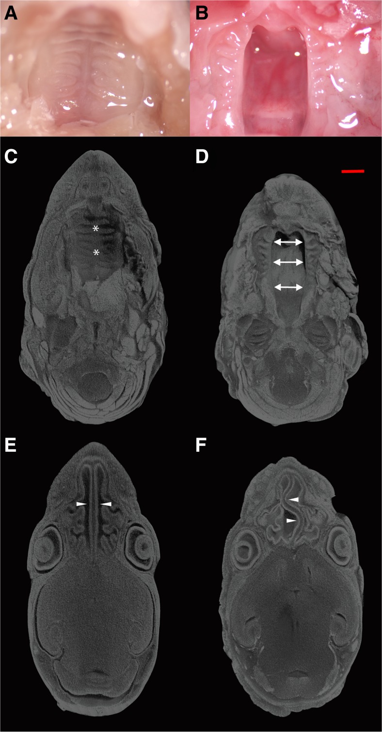
Gross specimen of wild type (WT; a) and CCN2 knockout (KO; b) palates in postnatal day 0 (P0) mice. MicroCT analysis of palate (top, c and d) and nasal septum/cavity (bottom, e and f) in (P0) heads from WT; (c and e) and KO; (d and f) mice. Specimens were stained with phosphotungstic acid to allow visualization and volumetric reconstruction of craniofacial tissues. The palate in WT mice (designated by * in c) is fully formed and fused, while remaining open in KO mice (designated by arrows in d). Nasal septum (arrowheads) and lateral wall of nasal cavity exhibit significant developmental deformities in KO (f) compared to WT (e) mice. Scale bar = 1 mm. (n = 38 WT and KO mice)

